청소년 methamphetamine 중독 1례
A case of methamphetamine intoxication in an adolescent
Article information

Trans Abstract
With the age of exposure to illegal substances decreasing and abuse of drugs such as methamphetamine increasing, substance abuse is no longer limited to adults. We report a Korean case of a 17-year-old girl with acute methamphetamine poisoning. The girl visited the emergency department for vomiting and loss of consciousness, with needle marks found on both arms. QT prolongation was confirmed on the initial electrocardiogram, so that we suspected drug addiction and proceeded with toxicologic tests. A lethal dose of methamphetamine was confirmed. We discontinued QT prolonging drugs, and closely monitored the girl in the pediatric emergency intensive care unit until the QT prolongation was resolved. This case highlights the recognition of pediatric methamphetamine poisoning in emergency departments.
Introduction
Methamphetamine, a methylated analog of amphetamine, shows a sympathomimetic toxidrome that is prominent in the central nervous and cardiovascular systems, such as agitation, hallucination, mydriasis, hyperthermia, hypertension, and tachycardia1,2). Despite the potential for poor outcomes, there has been a paucity of literature on methamphetamine toxicity in adolescents. A United States study shows that in 23 adolescents aged 12-18 years, 21 had tachycardia or hypertension, 11 mydriasis, and 6 gastrointestinal (GI) symptoms without a mortality3). A Turkish study shows that in 10 adolescents aged 14-17 years, 8 had tachycardia, 7 neurologic symptoms such as hallucination, and 5 GI symptoms without a mortality4). We report a Korean adolescent who self-injected methamphetamine to highlight the recognition of therapeutic principles of methamphetamine poisoning in adolescents with acute poisoning who visit emergency departments (EDs). This study was approved by the institutional review board of Seoul National University Hospital (IRB no. 2207-076-1339).
Case
A 17-year-old girl with major depressive disorder visited the ED with vomiting, myoclonus, and altered mentality in June 2022. When paramedics arrived at the scene, the girl could not communicate owing to decreased consciousness with bloody vomitus around her and several injection marks on both arms. Text messages on her cell phone suggested that she had met an unknown person in an unfamiliar place, and self-injected the drug.
The initial vital signs were as follows: blood pressure, 113/66 mmHg; heart rate, 112 beats/minute; respiratory rate, 20 breaths/minute; temperature, 38°C; oxygen saturation, 99% on room air; and an AVPU scale of V. Given that she complained of a headache and was agitated, we injected intramuscularly 4 mg of lorazepam and 5 mg of haloperidol. In addition, to investigate the drug that she administered on her arms, her blood and urine samples were sent to the toxicology laboratory. Although she had a fever, headache, and decreased consciousness, we did not perform lumbar puncture because she recovered soon. Fever was also improved without use of antipyretics or cooling.
QT prolongation was confirmed on the electrocardiogram (ECG) on arrival at the ED (Fig. 1). However, values of electrolytes and cardiac enzymes were within the normal ranges (Table 1). Brain computed tomography showed no intracranial hemorrhage. Regarding the bloody vomitus, we found an absence of active bleeding and GI perforation or ischemia on an abdominopelvic computed tomography. Consequently, the bleeding was presumed to come from Mallory-Weiss tear or methamphetamine per se. We decided to maintain conservative therapy using esomeprazole to reduce rebleeding, and administered intravenous ceftriaxone as a prophylactic antibiotic. After an overnight observation in the ED, she was referred for psychiatric consultation on day 2. Her toxicologic findings showed that the concentration of methamphetamine was as high as the reported postmortem blood concentration (Table 2)5,6). The psychiatrist confirmed that the risk of self-harm could not be excluded. Because close monitoring for QTc interval and hematemesis was necessary, she was hospitalized in the pediatric emergency intensive care unit.
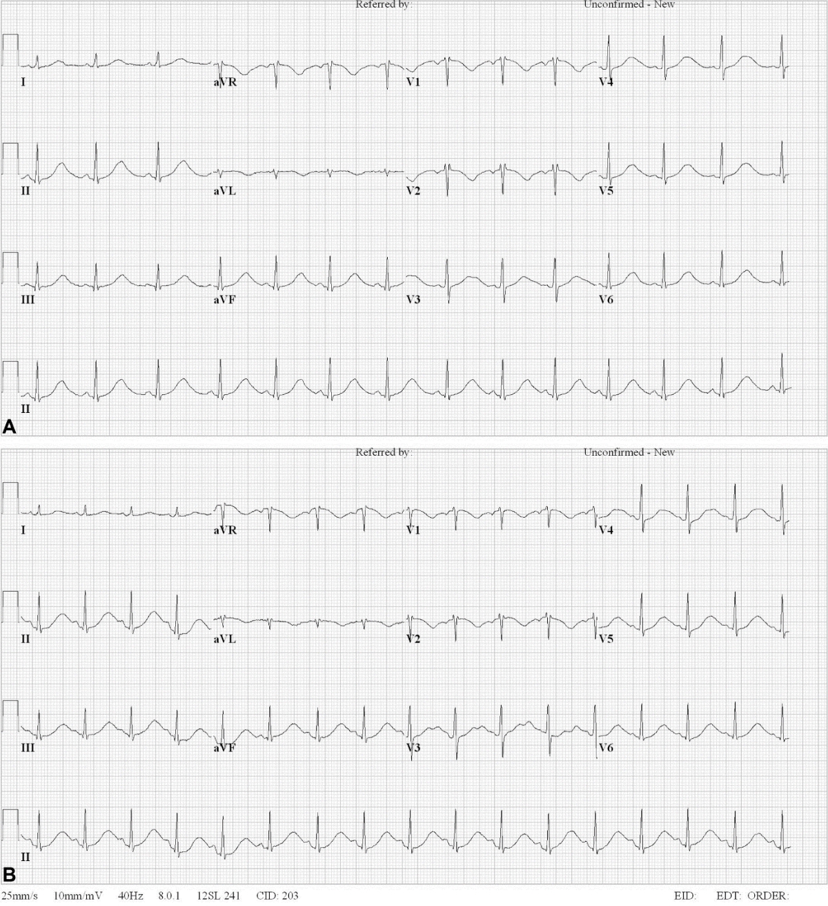
The changes in QTc interval on the electrocardiograms. It was 604 ms at the time of the visit (A) and 641 ms 6 hours after the visit (B).

Laboratory findings on arrival at the emergency department

Analysis of plasma concentrations of suspected substances
In the intensive care unit, the girl was alert with the agitation improved, and became able to provide the detailed medical history. She used to consume alcoholic beverages about twice a week, and on the day of the ED visit, she had 5 bottles of such beverages at dawn and returned home. She had also taken 0.4 g of methamphetamine, 2/3 of which was administered to the right arm and 1/3 to the left arm. At the time of the first use of methamphetamine 2 months ago, she collapsed from dizziness but did not visit any ED.
We continued intravenous fluid therapy and attempted to normalize QTc interval by regularly checking blood electrolyte concentrations and if needed, by supplying some electrolytes. QT prolongation disappeared on day 3, and she was transferred to the ward. There was no additional hematemesis during the hospital stay, so an endoscopy was not performed.
During the hospital stay, she underwent daily psychiatric interviews, and was advised to adjust psychiatric medications for persistent irritable mood and aggressive behavior. The girl was discharged uneventfully on day 8.
Discussion
We report a case of acute methamphetamine poisoning of a 17-year-old girl in Korea. This case highlights the recognition of pediatric methamphetamine poisoning in EDs by sharing information on toxidrome, diagnosis, and treatment process.
Pediatric patients with methamphetamine poisoning tend to have more prominent GI symptoms compared to adults. According to the U.S. study, 26.3% of patients younger than 18 years complained of GI symptoms, mainly nausea and vomiting, but 1 diarrhea, 1 hematemesis, and 1 hematochezia were also reported3). In the Turkish study, 50% of patients younger than 18 years showed GI symptoms, such as nausea and stomachache4). Methamphetamine use was also associated with GI ulcers, perforation, and bleeding in younger age groups compared to non-use patients7). A 7-year study of patients in a single ED in Korea who were positive for methamphetamine from a drug screening test showed that their age ranged from 21 to 84 years with a mean age of 37 years8). In that study, most patients reported neurologic symptoms, such as mental change, drowsiness or hallucinations. In a 3-year single ED study in Korea, 12 patients with a median age of 42 years were suspected to use methamphetamine on urine toxicologic screening tests, and reported tachycardia, mental change, agitation, and psychosis as major symptoms9).
It is important to be aware of the symptoms related to acute poisoning, and to conduct ECG after arrival at EDs. ECG should be monitored during the use of antipsychotics, such as haloperidol. In a previous study, antipsychotics have no fatal adverse effects3). Moreover, the presence of injection marks on the case patient’s arms could reveal the drug abuse. In the abovementioned Korean study, injection marks were more frequently found in the methamphetamine-true positive group than in the false positive group9). Hence, we also emphasize the physical examinations for patients suspected of acute poisoning.
With the recent decrease in the age of exposure to illegal substances and the increase in the abuse of drugs, such as methamphetamine, substance abuse is no longer limited to adults. In EDs, we need to prepare for cases of poisoning by substances that can cause fatal or serious sequelae in children and adolescents.
Notes
Conflicts of interest
Jae Yun Jung has been an associate editor of the Pediatric Emergency Medicine Journal since 2014. He was not involved in the review process. Otherwise, no potential conflicts of interest relevant to this article were reported.
Funding sources
No funding source relevant to this article was reported.