


ņä£ ļĪĀ
ņŗĀĻ▓Įļ¬©ņäĖĒżņóģņØĆ ņøÉņŗ£ ņŗĀĻ▓ĮļŖźņäĀņäĖĒżņŚÉņä£ ĻĖ░ņøÉĒĢ£ ņĢģņä▒ņóģņ¢æņ£╝ļĪ£ņä£, ņĪ░ņ¦üĒĢÖņĀüņ£╝ļĪ£ ņŗĀĻ▓Į ļČäĒÖöļÅäņŚÉ ļö░ļØ╝ ņŗĀĻ▓Įļ¬©ņäĖĒżņóģ, ņŗĀĻ▓ĮņĀłņŗĀĻ▓Įļ¬©ņäĖĒżņóģ, ņŗĀĻ▓ĮņĀłņŗĀĻ▓Įņóģņ£╝ļĪ£ ļČäļźśļÉ£ļŗż[1]. ņØ┤ņóģņ¢æņØĆ ņåīņĢä Ļ│ĀĒśĢņĢö ņżæ ļćīņóģņ¢æņŚÉ ņØ┤ņ¢┤ ļæÉ ļ▓łņ¦ĖļĪ£ ĒØöĒĢśļ®░,5ņäĖ ļ»Ėļ¦ī ņåīņĢäņŚÉņä£ ļČĆņŗĀ ņóģĻ┤┤ļĪ£ ļ░£ĒśäĒĢśļŖö Ļ▓ĮņÜ░Ļ░Ć Ļ░Ćņן ĒØö ĒĢśļŗż[2]. ĒĢśņ¦Ćļ¦ī, ņøÉļ░£ ļ░Å ņĀäņØ┤ ļČĆņ£äņØś ļŗżņ¢æĒĢ£ ņ”Øņāüņ£╝ļĪ£ ņØĖĒĢ┤ ņ¦äļŗ©ņØ┤ ņ¢┤ļĀżņÜ┤ Ļ▓ĮņÜ░Ļ░Ć ļ¦Äņ£╝ļ»ĆļĪ£, ņŗĀĻ▓Įļ¬©ņäĖĒżņóģņØä ņŗ£ņé¼ĒĢśļŖö ļŗżņ¢æĒĢ£ ņ×äņāüņ”ØņāüņŚÉ ļīĆĒĢśņŚ¼ Ļ░Éļ│ä ņ¦äļŗ©ĒĢĀ ņłś ņ׳ņ¢┤ņĢ╝ ĒĢ£ļŗż. ļ│Ė ņĀĆņ×ÉļŖö 3ņŻ╝Ļ░ä ņ¦ĆņåŹĒĢ£ ņ¢æņĖĪ Ļ│ĀĻ┤ĆņĀł ļ░Å ļ│ĄļČĆņØś ĒåĄņ”Øņ£╝ļĪ£ ļ│ĖņøÉ ņØæĻĖēņŗżņŚÉ ļ░®ļ¼ĖĒĢśņŚ¼, Ēīīņóģņä▒ ļ╝ł ļ░Å Ļ│©ņłś ņĀäņØ┤ļź╝ļÅÖļ░śĒĢ£ ņŗĀĻ▓Įļ¬©ņäĖĒżņóģņ£╝ļĪ£ ņ¦äļŗ©ļÉ£ 3ņäĖ ļé©ņ×É ĒÖśņ×ÉņØś ņ”ØļĪĆļź╝ļ│┤Ļ│ĀĒĢśĻ│Āņ×É ĒĢ£ļŗż. ļśÉĒĢ£, ņŗĀĻ▓Įļ¬©ņäĖĒżņóģņØś ņ×äņāüņ”Øņāü ļ░Å ņ£ĀņĢäĻĖ░ ļ╣äņÖĖņāüņä▒ Ļ│ĀĻ┤ĆņĀł ĒåĄņ”ØņØś Ļ░Éļ│ä ņ¦äļŗ©ņØä ņżæņŗ¼ņ£╝ļĪ£ Ļ░äļץĒĢśĻ▓ī Ļ│Āņ░░ĒĢśĻ│Āņ×É ĒĢ£ļŗż.
ņ”Ø ļĪĆ
3ņäĖ ļé©ņ×É ĒÖśņ×ÉĻ░Ć 3ņŻ╝Ļ░ä ņ¦ĆņåŹļÉ£ ņ¢æņĖĪ Ļ│ĀĻ┤ĆņĀł ļ░Å ļ│ĄļČĆņØśĒåĄņ”ØņØä ņŻ╝ņåīļĪ£ ļ│ĖņøÉ ņØæĻĖēņŗżņØä ļ░®ļ¼ĖĒ¢łļŗż. ĒÖśņ×ÉļŖö ļ│ĖņøÉ ļ░®ļ¼Ė 3ņŻ╝ ņĀä, ņÜ░ņĖĪ Ļ│ĀĻ┤ĆņĀł ļ░Å ļ│ĄļČĆ ĒåĄņ”ØņØ┤ ļ░£ņāØĒĢśņŚ¼ ņÖĖļČĆļ│æņøÉņŚÉņ×ģņøÉĒ¢łļŗż. ļŗ╣ņŗ£ ļ│ĄļČĆ ņ╗┤Ēō©Ēä░ļŗ©ņĖĄņ┤¼ņśü ĒīÉļÅģ Ļ▓░Ļ│╝ ĒŖ╣ņØ┤ņåīĻ▓¼ņØ┤ ņŚåņŚłņ¦Ćļ¦ī, Ļ│©ļ░ś ņ×ÉĻĖ░Ļ│Ąļ¬ģņśüņāüņŚÉņä£ ņ¢æņĖĪ Ļ│©ļ░ś ļ░Å ļīĆĒć┤Ļ│©ĻĘ╝ņ£äļČĆņØś ļŗżļ░£ņä▒ Ļ│©ļ│æņåīļź╝ ĻĘ╝Ļ▒░ļĪ£ ĻĖēņä▒ Ļ│©ņłśņŚ╝ņ£╝ļĪ£ ņ¦äļŗ©ļÉśņŚłļŗż. ņØ┤Ēøä ĒĢŁņāØņĀ£ ņ╣śļŻīņŚÉļÅä ļČłĻĄ¼ĒĢśĻ│Ā ņóīņĖĪ Ļ│ĀĻ┤ĆņĀłņØś ĒåĄņ”ØĻ│╝ ļ░£ņŚ┤ņØ┤ ļÅÖļ░śĒĢśļ®░ ņ”ØņāüņØ┤ ņĢģĒÖöĒ¢łļŗż. ņÖĖļČĆļ│æņøÉņŚÉņä£ ļŗżņŗ£ ņŗ£Ē¢ēĒĢ£ ņ×ÉĻĖ░Ļ│Ąļ¬ģņśüņāüņŚÉņä£ ņÜ░ņĖĪ Ļ│ĀĻ┤ĆņĀłņØś ĒÖ£ņĢĪ(synovial fluid) ņ”ØĻ░Ć ļ░Å ĒÖ£ļ¦ēņØś ņĪ░ņśüņ”ØĻ░Ģ ņåīĻ▓¼ņØä ļ│┤ņśĆņ£╝ļ®░, ĒĢ┤ļŗ╣ļ│æņøÉ ņØśļŻīņ¦äņØĆ ĻĖēņä▒ Ļ│©ņłśņŚ╝ņŚÉ ĒÖöļåŹĻ┤ĆņĀłņŚ╝ņØ┤ ļÅÖļ░śĒĢ£ Ļ▓āņ£╝ļĪ£ ņ¦äļŗ©Ē¢łļŗż. ĒÖśņ×ÉņØś ļČĆļ¬©ļŖö ņĢģĒÖöĒĢśļŖö ņ”ØņāüņŚÉ ļīĆĒĢ£ ņČöĻ░ĆņĀüņØĖ ņ¦äļŻīļź╝ ņ£äĒĢ┤ ļ│ĖņøÉņØä ļ░®ļ¼ĖĒ¢łļŗż.
ļ│ĖņøÉ ļ░®ļ¼Ė ļŗ╣ņŗ£ ņØśņŗØņØĆ ļ¬ģļŻīĒ¢łĻ│Ā, ĒÖ£ļĀźņ¦ĢĒøäļŖö ĒśłņĢĢ109/74 mmHg, ĒśĖĒØĪņłś 40ĒÜī/ļČä, ņŗ¼ņןļ░ĢļÅÖņłś 149ĒÜī/ļČä,ņ▓┤ņś© 38.1ŃĆéC, ņé░ņåīĒżĒÖöļÅä 100%ņśĆļŗż. ņÖĖļČĆļ│æņøÉ ņ×ģņøÉ ļŗ╣ņŗ£ļ│┤ļŗż Ļ│ĀĻ┤ĆņĀł ĒåĄņ”ØņØĆ ļ╣äņŖĘĒĢ£ ņĀĢļÅäņśĆņ¦Ćļ¦ī ļ│ĄĒåĄņØĆ ņĢģĒÖöļÉ£ ņāüĒā£ņśĆļŗż. ņŗĀņ▓┤Ļ▓Ćņé¼ņŚÉņä£ ļ│ĄļČĆ ņĢĢĒåĄ ļ░Å Ļ░äļ╣äņןļ╣äļīĆļŖö ņŚåņŚłņ¦Ćļ¦ī,ņÜ░ņĖĪ ļäōņĀüļŗżļ”¼ ņĢĢĒåĄĻ│╝ ņ¢æņĖĪ Ļ│ĀĻ┤ĆņĀłņØś ņÜ┤ļÅÖ ļ▓öņ£ä ņĀ£ĒĢ£ņØ┤ Ļ┤Ćņ░░ļÉśņŚłņ£╝ļ®░ ņ▓┤ņżæļČĆĒĢśļŖö ļČłĻ░ĆļŖźĒ¢łļŗż. ļ░£ņĀü, ļČĆņóģ, ļ®ŹņØĆ ļÅÖļ░śĒĢśņ¦Ć ņĢŖņĢśņ£╝ļ®░, ĒśĖĒØĪĻ│żļ×Ć ļ░Å ĻĄŁņåī ņŗĀĻ▓ĮĒĢÖņĀü Ļ▓░ņåÉņØĆ ņŚåņŚłļŗż.
ĒśłņĢĪĻ▓Ćņé¼ņŚÉņä£ ļ░▒ĒśłĻĄ¼ 7,300/╬╝L, Ēśłņāēņåī 7.9 g/dL, ĒśłņåīĒīÉ 310,000/╬╝L, ņĀüĒśłĻĄ¼ņ╣©Ļ░ĢņåŹļÅä 47 mm/h (ņ░ĖĻ│Āņ╣ś:0-9 mm/h) ĒöäļĪ£ĒŖĖļĪ¼ļ╣łņŗ£Ļ░ä 13.4ņ┤ł(ņ░ĖĻ│Āņ╣ś: 10-13ņ┤ł)ņśĆņ£╝ļ®░, ņĢäņŖżĒīīļź┤ĒģīņØ┤ĒŖĖņĢäļ»ĖļģĖņĀäļŗ¼ĒÜ©ņåī 44 IU/L, ņĢīļØ╝ļŗīņĢäļ»ĖļģĖņĀäļŗ¼ĒÜ©ņåī 24 IU/L, Ēü¼ļĀłņĢäĒŗ┤ņØĖņé░ĒÖöĒÜ©ņåī 69 IU/L (ņ░ĖĻ│Āņ╣ś: 50ŌĆō250 IU/L), ņĀ¢ņé░ĒāłņłśņåīĒÜ©ņåī 690 IU/L (ņ░ĖĻ│Āņ╣ś: 120ŌĆō250 IU/L), C-ļ░śņØæļŗ©ļ░▒ņ¦ł 18.3 mg/dL (ņ░ĖĻ│Āņ╣ś: 0ŌĆō0.6 mg/dL)ņśĆļŗż. ņĀäĒĢ┤ņ¦ł, Ēśłļŗ╣, Ēü¼ļĀłņĢäĒŗ░ļŗī, ņÜöĻ▓Ćņé¼ļŖö ņĀĢņāüņØ┤ņŚłļŗż.
ĒØēļČĆ, ļ│ĄļČĆ ļ░Å Ļ│ĀĻ┤ĆņĀł XņäĀĻ▓Ćņé¼ņŚÉņä£ ņĀĢņāü ņåīĻ▓¼ņØä ļ│┤ņśĆļŗż.ņÖĖļČĆļ│æņøÉ ņ×ÉĻĖ░Ļ│Ąļ¬ģņśüņāüņŚÉņä£ ņĀ£ĻĖ░ļÉ£ ĒÖöļåŹĻ┤ĆņĀłņŚ╝ ņåīĻ▓¼ņØä ĒÅēĻ░ĆĒĢśĻ│Āņ×É Ļ│ĀĻ┤ĆņĀł Ļ┤ĆņĀłņ▓£ņ×Éļź╝ ņŗ£Ē¢ēĒ¢łĻ│Ā, ņØ┤ļź╝ ĒåĄĒĢ┤ ņ▒äņĘ©ĒĢ£ĒÖ£ņĢĪ ļČäņäØĻ▓Ćņé¼ Ļ▓░Ļ│╝, ļ░▒ĒśłĻĄ¼ 450/╬╝L (ņżæņä▒ĻĄ¼ 53%)ņśĆļŗż.
ņśüņāüņØśĒĢÖĻ│╝ ņØśņé¼Ļ░Ć ņÖĖļČĆļ│æņøÉ ņ×ÉĻĖ░Ļ│Ąļ¬ģņśüņāü ļ░Å ņ╗┤Ēō©Ēä░ļŗ©ņĖĄņ┤¼ņśüņØä ļŗżņŗ£ ĒīÉļÅģĒĢ£ Ļ▓░Ļ│╝, ņĀäņ×ÉņŚÉņä£ ĒÖĢņØĖļÉ£ ļŗżļ░£ņä▒ Ļ│©ļ│æņåīĻ░Ć ĻĖēņä▒ Ļ│©ņłśņŚ╝ļ│┤ļŗż Ļ│©ņłś ņĀäņØ┤ņĢö ļśÉļŖö ĒśłņĢĪņĢöņØä ņŗ£ņé¼ĒĢ£ļŗżļŖö ņåīĻ▓¼ņØ┤, Ēøäņ×ÉņŚÉņä£ļŖö ņÖĖļČĆļ│æņøÉ ĒīÉļÅģņŚÉņä£ ĒÖĢņØĖļÉśņ¦Ć ņĢŖņØĆ ņóīņĖĪ ļČĆņŗĀ ļ░Å ļīĆņóģļ¦źņŻ╝ņ£äļ”╝ĒöäņĀłĻ│╝ ņ£ĄĒĢ®ļÉ£ ņóģĻ┤┤Ļ░Ć Ļ░üĻ░üĒÖĢņØĖļÉśņŚłļŗż. ņØśļŻīņ¦äņØĆ ņĪ░ņĀłļÉśņ¦Ć ņĢŖļŖö ļŗżļ░£ņä▒ ĒåĄņ”ØĻ│╝ ļ╣łĒśł, ņĀ¢ņé░ĒāłņłśņåīĒÜ©ņåī ņ”ØĻ░Ć, ļ╣äĻ░ÉņŚ╝ņä▒ ņ¦łĒÖśņØä ņŗ£ņé¼ĒĢśļŖö ĒÖ£ņĢĪ ļČäņäØĻ▓Ćņé¼ Ļ▓░Ļ│╝, ņÖĖļČĆļ│æņøÉ ņśüņāüĻ▓Ćņé¼ ņ×¼ĒīÉļÅģ Ļ▓░Ļ│╝ļź╝ ĻĘ╝Ļ▒░ļĪ£,ņŗĀĻ▓Įļ¬©ņäĖĒżņóģ ļśÉļŖö ļ░▒Ēśłļ│æņØś Ļ░ĆļŖźņä▒ņØä ņØśņŗ¼Ē¢łļŗż. ņØ┤ņŚÉ ņŗĀĻ▓Įļ¬©ņäĖĒżņóģ Ļ░ĆļŖźņä▒ņØä ĒÅēĻ░ĆĒĢśĻ│Āņ×É ļ│ĄļČĆ ņ╗┤Ēō©Ēä░ļŗ©ņĖĄņ┤¼ņśüņØä ņŗ£Ē¢ēĒĢ£ Ļ▓░Ļ│╝, ņóīņĖĪ ļČĆņŗĀ ļ░Å ļīĆļÅÖļ¦źņŻ╝ņ£äļ”╝ĒöäņĀłĻ│╝ ņ£ĄĒĢ®ļÉ£ ņóģĻ┤┤Ļ░Ć ņ×¼ĒÖĢņØĖļÉśņŚłļŗż(Fig. 1). ņØśļŻīņ¦äņØĆ ņØ┤ ņóģĻ┤┤Ļ░Ć ņŗĀĻ▓Įļ¬©ņäĖĒżņóģņØ╝ Ļ░ĆļŖźņä▒ņØ┤ ļåÆļŗżĻ│Ā ļ│┤Ļ│Ā, ņåīņĢäņ▓Łņåīļģä ņóģņ¢æĒśłņĢĪĻ│╝ ņ×ģņøÉņ╣śļŻīļź╝ Ļ▓░ņĀĢĒ¢łļŗż.
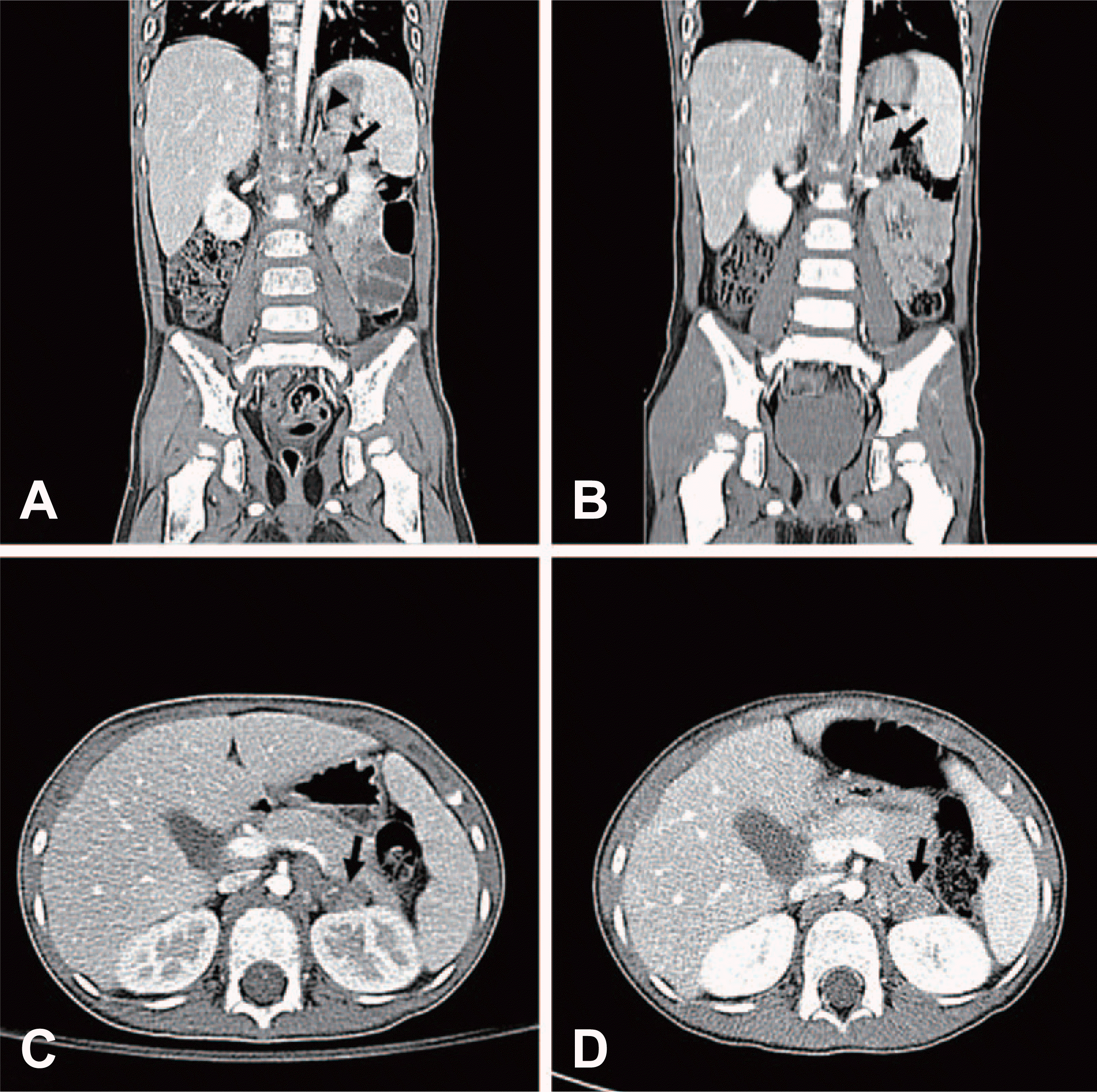
Fig.┬Ā1.
The results of computed tomography scan performed at our emergency department (A and C) and at another hospital (B and D), showing a mass indicating neuroblastoma. A contrast-enhanced mass in the left para-aortic area, approximately 20├Ś30 mm in size, is marked by the black arrows, and the size and location are comparable between the results from the two institutions. This mass abuts the left adrenal gland indicated by the black arrowheads in A and B.
ņ×ģņøÉ Ēøä, 24ņŗ£Ļ░ä ņåīļ│ĆĻ▓Ćņé¼ņŚÉņä£ homovanilic acid (HVA) ļ░Å vanillylmandelic acid (VMA)ļŖö Ļ░üĻ░ü 28.4 mg/d (ņ░ĖĻ│Āņ╣ś: 8.8 mg/d), 6.2 mg/d (ņ░ĖĻ│Āņ╣ś: 8.0 mg/d)ļĪ£ ĒÖĢņØĖļÉśņŚłĻ│Ā, ĒśłņĢĪĻ▓Ćņé¼ņŚÉņä£ ĒÄśļ”¼Ēŗ┤ 757.3 ng/mL (ņ░ĖĻ│Āņ╣ś: 20-320 ng/mL), ļē┤ļ¤░ĒŖ╣ņØ┤ņŚÉļåĆļØ╝ņĢäņĀ£(neuron-specific enolase) 98.5 ng/mL (ņ░ĖĻ│Āņ╣ś:4.7-14.7 ng/mL)ņśĆņ£╝ļ®░, ņØæĻĖēņŗżņŚÉņä£ ņŗ£Ē¢ēĒĢ£ ĒśłņĢĪ ļ░Å ĒÖ£ņĢĪ ļ░░ņ¢æĻ▓Ćņé¼ņŚÉņä£ ļÅÖņĀĢļÉ£ ņäĖĻĘĀņØĆ ņŚåņŚłļŗż. ņ¢æņĖĪ ņןĻ│©ļŖźņäĀņŚÉņä£ ņŗ£Ē¢ēĒĢ£ Ļ│©ņłśņāØĻ▓ĆņŚÉņä£, ņŗĀĻ▓Įļ¬©ņäĖĒżņóģņØ┤ ņ¦äļŗ©ļÉśņŚłļŗż.3,4-dihydroxy-6-18F-fluoro-phenylalanineņØä ņØ┤ņÜ®ĒĢ£ ņ¢æņĀäņ×Éļ░®ņČ£ļŗ©ņĖĄņ┤¼ņśüĻ│╝ 99 mTc-methylene diphosphonateņØä ņØ┤ņÜ®ĒĢ£ Ļ│©ņŖżņ║öņŚÉņä£ ņŗĀĻ▓Įļ¬©ņäĖĒżņóģņŚÉ ļÅÖļ░śĒĢ£ Ēīīņóģņä▒ ļ╝ł ļ░Å Ļ│©ņłś ņĀäņØ┤ ņåīĻ▓¼ņØä ļ│┤ņśĆļŗż(Fig. 2). ĒÖśņ×ÉļŖö International Neuroblastoma Staging SystemņŚÉļö░ļźĖ 4ĻĖ░, Children's Oncology Group Neuroblastoma Risk StratificationņŚÉ ļö░ļźĖ Ļ│Āņ£äĒŚśĻĄ░ņ£╝ļĪ£ ļČäļźśļÉśņŚłļŗż.ņØ┤Ēøä ņŗĀļ│┤Ļ░ĢĒÖöĒĢÖņÜöļ▓ĢņŚÉ ņØ┤ņ¢┤ņä£ ļ│ĄĻ░ĢĻ▓Į ļČĆņŗĀņĀłņĀ£ņłĀ(ņóīņĖĪ)ņØä ņŗ£Ē¢ēĒ¢łĻ│Ā, ņāØĻ▓ĆņØä ĒåĄĒĢ┤ Ļ│ĀļČäĒÖöļÅäļź╝ ļ│┤ņØ┤ļŖö ņŗĀĻ▓Įļ¬©ņäĖĒżņóģņ£╝ļĪ£ ĒÖĢņ¦äļÉśņŚłļŗż(Fig. 3). Ēśäņ×¼, ĒÖśņ×ÉļŖö ĒÖöĒĢÖņÜöļ▓Ģ Ēøä ņ×ÉĻ░Ć ņĪ░Ēśłļ¬©ņäĖĒż ņØ┤ņŗØ ņśłņĀĢņØ┤ļŗż.
Fig.┬Ā2.
Positron emission tomography (A), positron emission tomography/computed tomography (B), and bone scintigraphy (C: early; D: delayed) showing disseminated metastasis to the bone and bone marrow. Multifocal increased uptake is shown in the early images of the skull, and this finding is most apparent in the left parietal bone (arrows, A and B). Bone scintigraphy shows increased uptake in the bilateral pelvic bones and proximal femora.
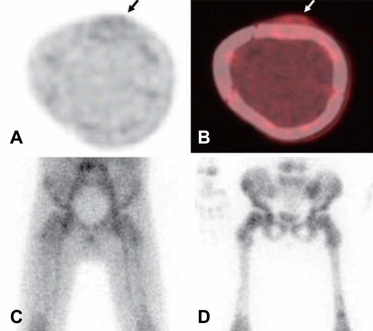
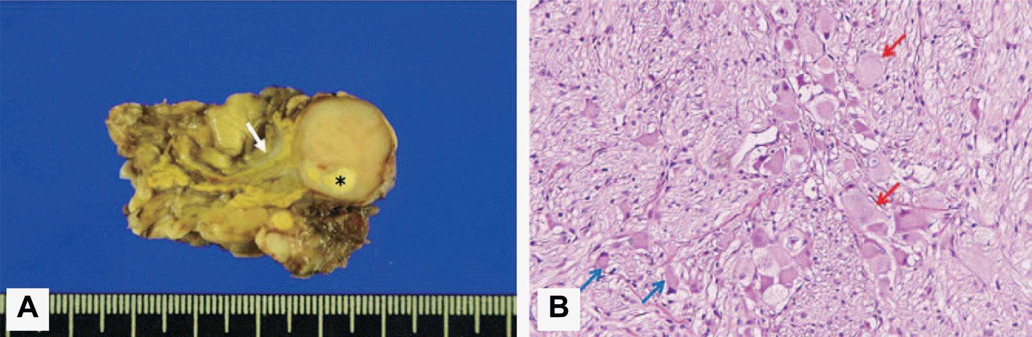
Fig.┬Ā3.
(A) Gross and (B) microscopic findings of the excised tumor after neoadjuvant chemotherapy. The gross specimen shows a well-demarcated, 19├Ś18├Ś16 mm-sized, round solid mass with whitish yellow cut surface and focal necrosis (asterisk, A). This mass abuts the left adrenal gland (white arrow, A). Most of the tumor is composed of Schwann cells. Foci of mature (red arrows, B) and maturing (blue arrows, B) ganglion cells are occasionally observed. There is no definite residual malignant component. Considering the neoadjuvant chemotherapy, overall findings suggest the diagnosis of neuroblastoma with extensive differentiation (H&E, ├Ś200).
Ļ│Ā ņ░░
ņŗĀĻ▓Įļ¬©ņäĖĒżņóģņØĆ 5ņäĖ ļ»Ėļ¦ī(ņżæņĢÖĻ░Æ 23Ļ░£ņøö)ņŚÉņä£ ĒśĖļ░£ĒĢśĻ│Ā, ņøÉļ░£ ļ░Å ņĀäņØ┤ ļČĆņ£ä(ļ╝ł, Ļ│©ņłś, ļ”╝ĒöäņĀł, Ļ░ä, Ēö╝ļČĆ)ņØś ļŗżņ¢æĒĢ£ ņ×äņāüņ”ØņāüņØä ļéśĒāĆļéĖļŗż[2]. ļČĆņŗĀ ņóģĻ┤┤ļĪ£ ļ░£ĒśäĒĢśļŖö Ļ▓ĮņÜ░Ļ░ĆĻ░Ćņן ĒØöĒĢśņ¦Ćļ¦ī(50%), ĻĄÉĻ░ÉņŗĀĻ▓ĮņĀłņØä ļö░ļØ╝ ņ¢┤ļŖÉ ļČĆņ£äņŚÉļÅäļ░£ņāØĒĢĀ ņłś ņ׳ļŗż[1]. ņóģĻ┤┤ ņ£äņ╣śņŚÉ ļö░ļØ╝, ĒśĖĒØĪĻ│żļ×Ć, ņāüļīĆņĀĢļ¦źņ”ØĒøäĻĄ░, ņé¼ņ¦Ćļ¦łļ╣ä, ĒśĖļäłņ”ØĒøäĻĄ░ņØ┤ ņ£Āļ░£ļÉĀ ņłś ņ׳ļŗż. ĒŖ╣Ē׳, Ļ│©ņłśņŚÉ ņĀäņØ┤ļÉ£ Ļ▓ĮņÜ░ ļ╣łĒśł ļ░Å ĒśłņåīĒīÉĻ░Éņåīņ”ØņØ┤ ļéśĒāĆļé£ļŗż[3]. ļ░£ņŚ┤, Ļ│ĀĒśłņĢĢ, ņäżņé¼ļź╝ ļÅÖļ░śĒĢśļŖö Ļ▓ĮņÜ░ņŚÉļŖö ĻĘ╝Ļ│©Ļ▓® Ļ░ÉņŚ╝, ņŗ¼ĒśłĻ┤Ć ļśÉļŖö ņ£äņןĻ┤Ć ņ¦łĒÖśĻ│╝ņØś Ļ░Éļ│äņØ┤ ņ¢┤ļĀĄļŗż. ļō£ļ¼╝ņ¦Ćļ¦ī ņĢłĻĄ¼Ļ░äļīĆĻ▓ĮļĀ©-ĻĘ╝Ļ░äļīĆĻ▓ĮļĀ© ņ”ØĒøäĻĄ░(opsoclonus-myoclonus syndrome)ņØ┤ ļ░£ņāØĒĢśĻĖ░ļÅä ĒĢ£ļŗż[4]. 5ņäĖ ļ»Ėļ¦ī ĒÖśņ×ÉĻ░Ć ņäżļ¬ģļÉśņ¦Ć ņĢŖļŖö ļŗżņ¢æĒĢ£ ļČĆņ£äņØś ĒåĄņ”Ø, ņŗĀĻ▓ĮĒĢÖņĀü ņ”Øņāü, ĒśĖĒØĪĻ│żļ×Ć, ļ░£ņŚ┤ ļ░Å Ļ│ĀĒśłņĢĢņØä ļ│┤ņØ╝ Ļ▓ĮņÜ░, Ļ░Éļ│ä ņ¦äļŗ©ņŚÉ ņŗĀĻ▓Įļ¬©ņäĖĒżņóģņØäĒżĒĢ©ĒĢ┤ņĢ╝ ĒĢ£ļŗż.
ļ│Ė ņ”ØļĪĆņÖĆ Ļ░ÖņØ┤ ļ╣äņÖĖņāüņä▒ Ļ│ĀĻ┤ĆņĀł ĒåĄņ”ØņØä ļ│┤ņØ┤ļŖö ņ£ĀņĢäĻĖ░ĒÖśņ×ÉņŚÉņä£ļŖö, ļŗżņ¢æĒĢ£ Ļ░Éļ│ä ņ¦äļŗ©ņØä Ļ│ĀļĀżĒĢ┤ņĢ╝ ĒĢ£ļŗż(Table 1).ņØ╝Ļ│╝ņä▒ Ļ│ĀĻ┤ĆņĀł ĒÖ£ļ¦ēņŚ╝ņØĆ 3ŌĆō8ņäĖ ĒÖśņ×ÉņŚÉņä£ ĒśĖļ░£ĒĢśĻ│Ā ļīĆĻ░£ļ░£ņŚ┤ ņŚåņØ┤ 3ņŻ╝ ņØ┤ļé┤ņŚÉ ĒśĖņĀäļÉśļ»ĆļĪ£[5], ļ│Ė ņ”ØļĪĆņÖĆ ļ¦×ņ¦Ć ņĢŖļŖöļŗż. ļĀłĻĘĖ-ņ╣╝ļ▓Ā-ĒÄśļź┤ĒģīņŖżļ│æ(LeggŌĆōCalv├®ŌĆōPerthes disease)ņØĆ 4ŌĆō9ņäĖ ĒÖśņ×ÉņŚÉņä£ Ļ│ĀĻ┤ĆņĀł ĒåĄņ”ØņØ┤ 3ņŻ╝ ņØ┤ņāü ņ¦Ć ņåŹĒĢśĻ│Ā ņĢĮ 10%ņŚÉņä£ ņ¢æņĖĪņä▒ņ£╝ļĪ£ ļéśĒāĆļéĀ ņłś ņ׳ņ¦Ćļ¦ī[6], ļ░£ņŚ┤ļ░Å ļŗżļ░£ņä▒ ĒåĄņ”ØņØä ļÅÖļ░śĒĢśņ¦ä ņĢŖļŖöļŗż. Ļ│©ņłśņŚ╝ņØ┤ļéś ĒÖöļåŹĻ┤ĆņĀłņŚ╝ņØĆ ļŗ©ņØ╝ ļČĆņ£äļź╝ ņ╣©ļ▓öĒĢśļŖö Ļ▓ĮņÜ░Ļ░Ć ĒØöĒĢśļ®░(>80%) [7], ĒÖ£ņĢĪļ░▒ĒśłĻĄ¼Ļ░Ć 50,000/╬╝L ņØ┤ņāüņØĖ Ļ▓ĮņÜ░Ļ░Ć ļīĆļČĆļČäņØ┤ļŗż[8,9]. ļ│Ė ņ”ØļĪĆņŚÉņä£ļŖö ņ▓┤ņżæļČĆĒĢśĻ░Ć ļČłĻ░ĆļŖźĒĢśņśĆĻ│Ā ļ╣ĀļźĖ ņĀüĒśłĻĄ¼ņ╣©Ļ░ĢņåŹļÅä(47 mm/h) ļ░Å ļåÆņØĆ C-ļ░śņØæļŗ©ļ░▒ņ¦ł(18.3 mg/dL)ņØä ļ│┤ņśĆļŖöļŹ░, ņØ┤ļŖö Caird ļō▒[9]ņØś ĻĖ░ņżĆņŚÉ ļö░ļź┤ļ®┤ ĒÖöļåŹĻ┤ĆņĀłņŚ╝ Ļ░ĆļŖźņä▒ņØ┤ ņĢĮ 83%ņŚÉ ĒĢ┤ļŗ╣ĒĢśļŖö ņāüĒÖ®ņØ┤ņŚłļŗż. ņØ┤ņŚÉ Ļ┤ĆņĀłņ▓£ņ×Éļź╝ ņŗ£Ē¢ēĒĢśņśĆņ¦Ćļ¦ī, ĒÖ£ņĢĪ ļ░▒ĒśłĻĄ¼ļŖö 450/╬╝L (ņżæņä▒ĻĄ¼ 53%)ņśĆļŗż.Ļ▓Ćņé¼ Ļ▓░Ļ│╝ ĒĢ┤ņäØņŚÉ Ļ▓Ćņé¼ ņĀä ĒĢŁņāØņĀ£ Ēł¼ņŚ¼ ņé¼ņŗżņØä Ļ│ĀļĀżĒĢ┤ņĢ╝ĒĢśĻ▓Āņ¦Ćļ¦ī, Ļ▓░Ļ│╝ņĀüņ£╝ļĪ£ ĒÖ£ņĢĪ ļ░░ņ¢æĻ▓Ćņé¼ņŚÉņä£ ņäĖĻĘĀņØĆ ļÅÖņĀĢļÉśņ¦Ć ņĢŖņĢśļŗż. Ļ▓īļŗżĻ░Ć, ļ│ĄĒåĄ ņĢģĒÖöļŖö ĻĘ╝Ļ│©Ļ▓® Ļ░ÉņŚ╝ņ£╝ļĪ£ ņäżļ¬ģĒĢśĻĖ░ ņ¢┤ļĀĄļŗż. ņåīņĢäĻĖ░ ĒŖ╣ļ░£Ļ┤ĆņĀłņŚ╝(juvenile idiopathic arthritis)ņØĆ ļīĆĻ░£ ĒåĄņ”ØņØś Ļ░ĢļÅäĻ░Ć ņĢĮĒĢśĻ│Ā 6ņŻ╝ ņØ┤ņāüņŚÉ Ļ▒Ėņ│Éņä£ņä£Ē׳ ļ░£ņāØĒĢśļŖö Ļ▓ĮņÜ░Ļ░Ć ļ¦Äļŗż[10]. ņØ┤ ņżæ ņåīņłśĻ┤ĆņĀłĒśĢņØĆ 2ŌĆō4ņäĖ ĒÖśņ×ÉņŚÉņä£ 4Ļ░£ ņØ┤ĒĢśņØś Ļ┤ĆņĀłņØä ņ╣©ļ▓ö(ņŻ╝ļĪ£ ļ¼┤ļ”Ä ļ░Å ļ░£ļ¬®)ĒĢśĻ│Ā ņĢäņ╣©Ļ▓Įņ¦üņØä ļÅÖļ░śĒĢśļŖöļŹ░[11], Ļ│ĀĻ┤ĆņĀł ļŗ©ļÅģņ£╝ļĪ£ ļ░£ņāØĒĢśĻ▒░ļéś ņ▓┤ņżæļČĆĒĢśĻ░Ć ļČłĻ░ĆļŖźĒĢ£ Ļ▓ĮņÜ░ļŖö ļō£ļ¼╝ļŗż[10]. ņäżļ¬ģļÉśņ¦Ć ņĢŖļŖöļŗżņ¢æĒĢ£ ļČĆņ£äņØś ĒåĄņ”ØņŚÉ ļ╣łĒśł ļ░Å ņĀ¢ņé░ĒāłņłśņåīĒÜ©ņåī ņ”ØĻ░ĆĻ░Ć ļÅÖļ░śĒĢ£ Ļ▓ĮņÜ░, ņŗĀĻ▓Įļ¬©ņäĖĒżņóģ ļ░Å ļ░▒Ēśłļ│æĻ│╝ Ļ░ÖņØĆ ņóģņ¢æņØä Ļ│ĀļĀżĒĢ┤ņĢ╝ ĒĢ£ļŗż[3]. Ļ│©ņ£ĪņóģĻ│╝ Ļ░ÖņØĆ ņøÉļ░£Ļ│©ņóģņ¢æļÅä Ļ│ĀļĀżĒĢ┤ņĢ╝ ĒĢśņ¦Ćļ¦ī,ņé¼ņČśĻĖ░ņŚÉ ĒśĖļ░£ĒĢśļ»ĆļĪ£[3], Ļ░ĆļŖźņä▒ņØ┤ ļ¢©ņ¢┤ņ¦äļŗż.
Table┬Ā1.
Differential diagnosis of non-traumatic limp pain in children
ņŗĀĻ▓Įļ¬©ņäĖĒżņóģņØś ņ¦äļŗ© ĻĖ░ļ▓ĢņØĆ ņåīļ│ĆĻ▓Ćņé¼, ņśüņāüĻ▓Ćņé¼, ņāØĻ▓Ć(ņøÉļ░£ņóģņ¢æ, Ļ│©ņłś)ņ£╝ļĪ£ ļéśļē£ļŗż. ņåīļ│ĆņØä ĒåĄĒĢśņŚ¼ ņ╣┤ĒģīņĮ£ņĢäļ»╝ ļīĆņé¼ņé░ļ¼╝ņØĖ HVA ļ░Å VMAļź╝ Ļ▓Ćņé¼ĒĢĀ ņłś ņ׳ļŗż. ņ╗┤Ēō©Ēä░ļŗ©ņĖĄņ┤¼ņśü ļ░Åņ×ÉĻĖ░Ļ│Ąļ¬ģņśüņāüņØä ĒåĄĒĢśņŚ¼, ņøÉļ░£ņóģņ¢æĻ│╝ ĒØēĻ░Ģ ļ░Å ļ│ĄĻ░Ģ ļé┤ ņĀäņØ┤ļź╝ĒÖĢņØĖĒĢĀ ņłś ņ׳ļŗż. ņóģĻ┤┤ļŖö ļ│ĄļČĆ ņżæņĢÖņäĀņØä ļäśĻ▒░ļéś ļé┤ļČĆņŚÉ Ļ┤┤ņé¼ļ░Å ņČ£ĒśłņØä ļÅÖļ░śĒĢśļŖö Ļ▓ĮņÜ░Ļ░Ć ĒØöĒĢśļŗż. ļČĆņŗĀ ņÖĖ ļśÉļŖö ņĀäņØ┤ņä▒ ņóģĻ┤┤Ļ░Ć ņØśņŗ¼ļÉśļŖö Ļ▓ĮņÜ░, 123I-metaiodobenzylguanidineņØä ņØ┤ņÜ®ĒĢ£ ņä¼Ļ┤æņĪ░ņśüņłĀ ļśÉļŖö ņ¢æņĀäņ×Éļ░®ņČ£ļŗ©ņĖĄņ┤¼ņśüĻ│╝ Ļ░ÖņØĆ ĻĖ░ļŖźņĀüņśüņāüĻ▓Ćņé¼ļź╝ ĒåĄĒĢśņŚ¼ ĻĄÉĻ░ÉņŗĀĻ▓Į ĒÖ£ņä▒ļÅäļź╝ ĒÖĢņØĖĒĢśļŖö Ļ▓āņØ┤ ņżæņÜöĒĢśļŗż[12]. Ļ│©ņłś Ļ▓Ćņ▓┤ņŚÉņä£ small round blue cellņØ┤ ļĪ£ņĀ£ĒŖĖ(rosette)ļź╝ ĒśĢņä▒ĒĢśļŖö Ļ▓āņØ┤ Ļ┤Ćņ░░ļÉśĻ│Ā ņåīļ│ĆņŚÉņä£ HVA ļ░Å VMA ļåŹļÅäĻ░Ć ņ”ØĻ░ĆĒĢ£ Ļ▓ĮņÜ░ņŚÉļŖö ņ¦äļŗ©ņØ┤ Ļ░ĆļŖźĒĢśļŗż[2]. ĒĢśņ¦Ćļ¦ī,ĒÖĢņ¦ä, ņśłĒøä ņśłņĖĪ ļ░Å ņ╣śļŻī Ļ│äĒÜŹ Ļ▓░ņĀĢņŚÉ ņ׳ņ¢┤ņä£ ņøÉļ░£ņóģņ¢æņØś ņāØĻ▓ĆņØĆ ĒĢäņłśņĀüņØ┤ļŗż. ņĀäņĢöņ£ĀņĀäņ×É MYCNņØś ņ”ØĒÅŁņØĆ ļåÆņØĆ ļ│æĻĖ░ļ░Å ļČłļ¤ēĒĢ£ ņśłĒøäņÖĆ ņŚ░Ļ┤ĆļÉśļ»ĆļĪ£, ņ£ĀņĀäņ×ÉĻ▓Ćņé¼ļź╝ ĒåĄĒĢśņŚ¼ ņØ┤ļź╝ ĒÖĢņØĖĒĢ┤ņĢ╝ ĒĢ£ļŗż[1].
ļ│Ė ņ”ØļĪĆņŚÉņä£ ņ¦äļŗ©ņØ┤ ņ¦ĆņŚ░ļÉ£ ņØ┤ņ£Āļź╝ ļŗżņØīĻ│╝ Ļ░ÖņØ┤ ņÜöņĢĮĒĢĀņłś ņ׳ļŗż. ņ▓½ņ¦Ė, ņÖĖļČĆļ│æņøÉ ņ×ģņøÉ ļŗ╣ņŗ£ Ļ│ĀĻ┤ĆņĀł ĒåĄņ”ØņØ┤ ļ│ĄĒåĄļ│┤ļŗż ņŗ¼Ē¢łĻĖ░ ļĢīļ¼ĖņŚÉ, ņĢģņä▒ņóģņ¢æņØä Ļ│ĀļĀżĒĢśņ¦Ć ļ¬╗ĒĢ£ ņāüĒā£ņŚÉņä£ ļ│ĄļČĆ ņ╗┤Ēō©Ēä░ļŗ©ņĖĄņ┤¼ņśüņØä ņŗ£Ē¢ēĒ¢łļŗżļŖö ņĀÉņØ┤ļŗż. ņ”ē, ņČ®ņłśņŚ╝Ļ│╝ Ļ░ÖņØĆ ĻĖēņä▒ļ│Ąņ”ØņØś Ļ░Éļ│ä ņ¦äļŗ©ņØ┤ ņ┤¼ņśü ļ¬®ņĀüņØ┤ņŚłĻĖ░ ļĢīļ¼ĖņŚÉ, ņ┤¼ņśüĒøäņŚÉļÅä ļČĆņŗĀ ņŻ╝ņ£äļź╝ ĒÖĢņØĖĒĢśņ¦Ć ļ¬╗Ē¢łņØä ņłś ņ׳ļŗż. ļæśņ¦Ė, ļŗ╣ņŗ£ĒśłņāēņåīĻ░Ć 9.6ŌĆō11.6 g/dLļĪ£ ļ│ĖņøÉ Ļ▓Ćņé¼ Ļ▓░Ļ│╝ņŚÉ ļ╣äĒĢ┤ ļåÆņĢśļŹś Ļ▓āļÅä, ņĢģņä▒ņóģņ¢æņØä Ļ│ĀļĀżĒĢśņ¦Ć ļ¬╗ĒĢ£ Ļ▓āņŚÉ ĻĖ░ņŚ¼Ē¢łņØä ņłś ņ׳ļŗż. ņģŗņ¦Ė, ņ×ÉĻĖ░Ļ│Ąļ¬ģņśüņāüņŚÉņä£ ļéśĒāĆļé£ ļŗżļ░£ņä▒ Ļ│©ļ│æņåīļź╝ ĻĖēņä▒Ļ│©ņłśņŚ╝ņ£╝ļĪ£ ļŗ©ņĀĢĒ¢łļŗżļŖö ņĀÉņØ┤ļŗż. Ļ│©ņłśņŚ╝ņØĆ ļīĆļČĆļČä ļŗ©ņØ╝ ļ│æņåīļĪ£ ļ░£ņāØĒĢśĻ│Ā ĒÖśņ×ÉĻ░Ć ĒĢŁņāØņĀ£ ņ╣śļŻīņŚÉ ļ░śņØæĒĢśņ¦Ć ņĢŖņĢśņ£╝ļ»ĆļĪ£, ņĢģņä▒ņóģņ¢æņØś ļ╝ł ļ░Å Ļ│©ņłś ņĀäņØ┤ļź╝ ņØśņŗ¼Ē¢łņ¢┤ņĢ╝ Ē¢łļŗż. ĒĢśņ¦Ćļ¦ī, Ļ│©ņŖżņ║öĻ│╝ Ļ░ÖņØĆ Ļ▓Ćņé¼ļŖö ņČöĻ░ĆļĪ£ ņŗ£Ē¢ēļÉśņ¦Ć ņĢŖņĢśļŗż.
ļ│Ė ņ”ØļĪĆņŚÉņä£, ņØśļŻīņ¦äņØĆ ņĪ░ņĀłļÉśņ¦Ć ņĢŖļŖö ļŗżļ░£ņä▒ ĒåĄņ”ØĻ│╝ ĒśłņĢĪĒĢÖņĀü ņØ┤ņāü ņåīĻ▓¼, ļ╣äĻ░ÉņŚ╝ņä▒ ņ¦łĒÖśņØä ņŗ£ņé¼ĒĢśļŖö ĒÖ£ņĢĪ ļČäņäØĻ▓Ćņé¼ Ļ▓░Ļ│╝, ņŗĀĻ▓Įļ¬©ņäĖĒżņóģ ļ░Å Ēīīņóģņä▒ ļ╝ł ļ░Å Ļ│©ņłś ņĀäņØ┤ļź╝ ņŗ£ņé¼ĒĢśļŖö ņÖĖļČĆļ│æņøÉ ņśüņāüĻ▓Ćņé¼ ņ×¼ĒīÉļÅģ Ļ▓░Ļ│╝ļź╝ ĻĘ╝Ļ▒░ļĪ£ ņŗĀĻ▓Įļ¬©ņäĖĒżņóģ Ļ░ĆļŖźņä▒ņØä Ļ│ĀļĀżĒ¢łļŗż. ņØ┤Ēøä ņ╗┤Ēō©Ēä░ļŗ©ņĖĄņ┤¼ņśü, ņåīļ│Ć HVA ļ░Å VMA Ļ▓Ćņé¼ļź╝ ĒåĄĒĢ┤ ņŗĀĻ▓Įļ¬©ņäĖĒżņóģņ£╝ļĪ£ ņ×ĀņĀĢ ņ¦äļŗ©Ē¢łĻ│Ā, ņøÉļ░£ņóģņ¢æ ļ░Å Ļ│©ņłśņØś ņāØĻ▓ĆņØä ĒåĄĒĢ┤ ņ¦äļŗ©ņØä ĒÖĢņØĖĒ¢łļŗż.ņŗĀĻ▓Įļ¬©ņäĖĒżņóģņØĆ ļŗżņ¢æĒĢ£ ņ×äņāüņ”ØņāüņØä ļéśĒāĆļé╝ ņłś ņ׳ļŗż. ĒŖ╣Ē׳,5ņäĖ ļ»Ėļ¦ī ĒÖśņ×ÉņŚÉņä£ ņäżļ¬ģļÉśņ¦Ć ņĢŖļŖö ļŗżņ¢æĒĢ£ ļČĆņ£äņØś ĒåĄņ”ØņŚÉļ╣łĒśł ļ░Å ņĀ¢ņé░ĒāłņłśņåīĒÜ©ņåī ņ”ØĻ░ĆĻ░Ć ļÅÖļ░śĒĢ£ Ļ▓ĮņÜ░, ņŗĀĻ▓Įļ¬©ņäĖĒżņóģņØä Ļ░Éļ│ä ņ¦äļŗ©ņ£╝ļĪ£ Ļ│ĀļĀżĒĢ┤ņĢ╝ ĒĢ£ļŗż.


